HONDA ELECTRONICS Ultrasound System
HONDA ELECTRONICS diagnostic ultrasound systems are highly evaluated not only in Japan but also in various other countries. Here, we introduce some of the strengths of Honda Electronics, which have been highly evaluated by many users.

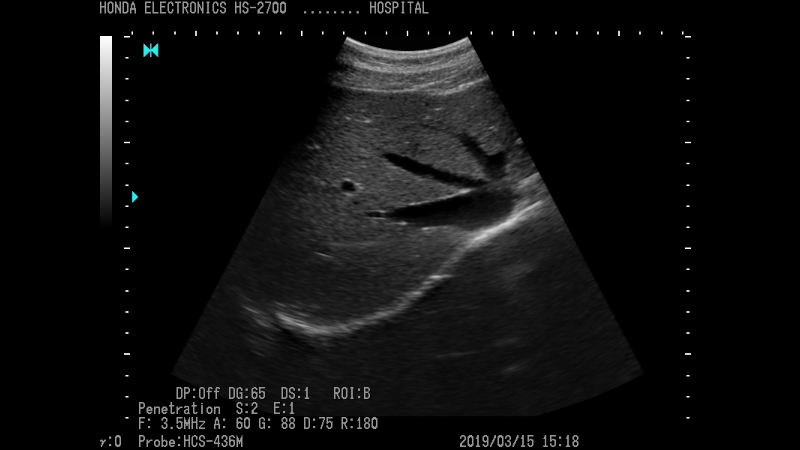
Medical Products
This section introduces products for human use for medical professionals.
We can help customers find ultrasound imaging systems and probes suitable for their use in catheterization, orthopedics, general internal medicine, obstetrics and gynecology, and other situations.
We can help customers find ultrasound imaging systems and probes suitable for their use in catheterization, orthopedics, general internal medicine, obstetrics and gynecology, and other situations.

Vetrinary Products
We can help customers find the right ultrasound imaging equipment and probes for their use scenarios, such as pregnancy appraisal and breeding, meat quality determination, and use with small animals.
-
Here, you can download catalogs and documents in PDF format.
Read more
-
Here are some frequently asked questions.
Read more
-

 About the list of discontinued models
Discontinued models are listed here.
Read more
-
For inquiries about our products, please contact us here.
Read more


